Figure 2
Arid3a regulates mesoderm differentiation in mouse embryonic stem cells
Haley O Tucker*, Melissa Popowski, Bum-kyu Lee, Vishwanath R Iyer and Cathy Rhee
Published: 07 September, 2017 | Volume 1 - Issue 1 | Pages: 052-062
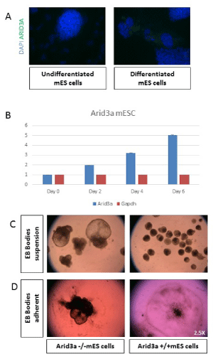
Figure 2:
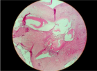
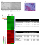
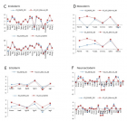
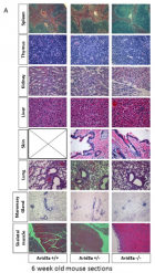
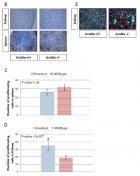
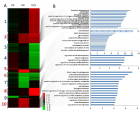
Arid3a increases expression and influences differentiation. (A) mES cells show an increase in Arid3a protein levels upon differentiation. mES cells were grown in chamber slides in the presence (undifferentiated) or absence (differentiated) of LIF for 3 days. Cells were stained for Arid3a expression (green), and nuclei were counterstained with DAPI (blue). (B) Arid3a +/+ mESC show an increase in Arid3a expression during differentiation induced through removal of stem cell factors compared to GAPDH expression. (C) Arid3a -/- mESC differentiate into embryoid bodies more rapidly compared to wildtype. Two different Arid3a-/- ES cell lines (Arid3a-/- ES-1 and Arid3a-/- ES-2) and Arid3a+/+ ES cells were grown in suspension culture without LIF to promote differentiation. Arid3a-/- ES formed CEBs more quickly, more often, and much larger then Arid3a+/+ ES. Images are at day 10 differentiation. 2.5x magnification. (C) Arid3a-/- mES do not differentiate correctly in adherent cultures. Two different Arid3a-/- cell lines and Arid3a+/+ mES cells were grown in suspension culture without LIF to promote formation of EBs for eight days. EBs were allowed to adhere to gelatin coated cell culture plates. Arid3a-/- mES cells did not appropriately adhere to the cell culture plate and appeared to incompletely differentiate. Images are at day 13 differentiation (2.5x magnification).
Read Full Article HTML DOI: 10.29328/journal.jsctt.1001005 Cite this Article Read Full Article PDF
More Images
Similar Articles
-
The Femoral Head of Patients with Hip Dysplasia is not as Osteogenic as Iliac Crest Bone LocationPhilippe Hernigou*,Yasuhiro Homma,Arnaud Dubory,Jacques Pariat,Damien Potage,Charles Henri Flouzat Lachaniette,Nathalie Chevallier,Helene Rouard . The Femoral Head of Patients with Hip Dysplasia is not as Osteogenic as Iliac Crest Bone Location. . 2017 doi: 10.29328/journal.jsctt.1001001; 1: 001-007
-
Rhabdomyoblasts in Pediatric Tumors: A Review with Emphasis on their Diagnostic UtilityGiuseppe Angelico*,Eliana Piombino,Giuseppe Broggi,Fabio Motta,Saveria Spadola. Rhabdomyoblasts in Pediatric Tumors: A Review with Emphasis on their Diagnostic Utility . . 2017 doi: 10.29328/journal.jsctt.1001002; 1: 008-016
-
Surgical Implantation of Stem Cells in Heart Failure Patients due to Idiophatic CardiomyopathyBenetti Federico*,Natalia Scialacomo,Enrique Mariani,Luis Geffner,Bruno Benetti Eng1,Daniel Brusich,Yan Duarte. Surgical Implantation of Stem Cells in Heart Failure Patients due to Idiophatic Cardiomyopathy. . 2017 doi: 10.29328/journal.jsctt.1001003; 1: 017-027
-
Enhancing adipose stem cell chondrogenesis: A study on the roles of dexamethasone, transforming growth factor β3 and ascorbate supplements and their combinationBernard J Van Wie*,Arshan Nazempour##,Chrystal R Quisenberry##,Nehal I Abu-Lail. Enhancing adipose stem cell chondrogenesis: A study on the roles of dexamethasone, transforming growth factor β3 and ascorbate supplements and their combination. . 2017 doi: 10.29328/journal.jsctt.1001004; 1: 028-051
-
Arid3a regulates mesoderm differentiation in mouse embryonic stem cellsHaley O Tucker*,Melissa Popowski,Bum-kyu Lee,Vishwanath R Iyer,Cathy Rhee. Arid3a regulates mesoderm differentiation in mouse embryonic stem cells. . 2017 doi: 10.29328/journal.jsctt.1001005; 1: 052-062
-
Nicotinamide as a treatment option of Age-Related Macular DegenerationChristine Skerka*,Yuchen Lin,Peter F. Zipfel. Nicotinamide as a treatment option of Age-Related Macular Degeneration. . 2017 doi: 10.29328/journal.jsctt.1001006; 1: 063-065
-
Intraepidermal Injections of Autologous Epidermal Cell Suspension: A new promising approach to Dermatological Disorders. Preliminary StudyFabio Rinaldi*,Elisa Borsani,Luigi Fabrizio Rodella,Elisabetta Sorbellini,Rita Rezzani,Daniela Pinto,Barbara Marzani,Giovanna Tabellini,Mariangela Rucco. Intraepidermal Injections of Autologous Epidermal Cell Suspension: A new promising approach to Dermatological Disorders. Preliminary Study. . 2017 doi: 10.29328/journal.jsctt.1001007; 1: 066-070
-
Stemness of Mesenchymal Stem CellsTong Ming Liu*. Stemness of Mesenchymal Stem Cells. . 2017 doi: 10.29328/journal.jsctt.1001008; 1: 071-073
-
Stem cells in heart failure some considerationsFederico Benetti*,Natalia Scialacomo,Bruno Benetti. Stem cells in heart failure some considerations. . 2018 doi: 10.29328/journal.jsctt.1001009; 2: 001-003
-
Stem cells in patients with heart failure experienceBenetti Federico*,Natalia Scialacomo. Stem cells in patients with heart failure experience. . 2018 doi: 10.29328/journal.jsctt.1001010; 2: 004-014
Recently Viewed
-
Sinonasal Myxoma Extending into the Orbit in a 4-Year Old: A Case PresentationJulian A Purrinos*, Ramzi Younis. Sinonasal Myxoma Extending into the Orbit in a 4-Year Old: A Case Presentation. Arch Case Rep. 2024: doi: 10.29328/journal.acr.1001099; 8: 075-077
-
Improvement of the Cognitive Abilities in a Chronic Generalized Anxiety Disorder and Moderate Depression Case using a Novel Integrated Approach: The Cognitome ProgramMohita Shrivastava*. Improvement of the Cognitive Abilities in a Chronic Generalized Anxiety Disorder and Moderate Depression Case using a Novel Integrated Approach: The Cognitome Program. J Neurosci Neurol Disord. 2024: doi: 10.29328/journal.jnnd.1001100; 8: 069-089
-
Neuroprotective Effect of 7,8-dihydroxyflavone in a Mouse Model of HIV-Associated Neurocognitive Disorder (HAND)Tapas K Makar, Joseph Bryant, Bosung Shim, Kaspar Keledjian, Harry Davis, Manik Ghosh, Ajay Koirala, Ishani Ghosh, Shreya Makar, Alonso Heredia, Malcolm Lane, J Marc Simard, Robert C Gallo, Volodymyr Gerzanich*, Istvan Merchenthaler*. Neuroprotective Effect of 7,8-dihydroxyflavone in a Mouse Model of HIV-Associated Neurocognitive Disorder (HAND). J Neurosci Neurol Disord. 2024: doi: 10.29328/journal.jnnd.1001101; 8: 090-105
-
Adult Neurogenesis: A Review of Current Perspectives and Implications for Neuroscience ResearchAlex, Gideon S*,Olanrewaju Oluwaseun Oke,Joy Wilberforce Ekokojde,Tolulope Judah Gbayisomore,Martina C. Anene-Ogbe,Farounbi Glory,Joshua Ayodele Yusuf. Adult Neurogenesis: A Review of Current Perspectives and Implications for Neuroscience Research. J Neurosci Neurol Disord. 2024: doi: 10.29328/journal.jnnd.1001102; 8: 106-114
-
Analysis of Psychological and Physiological Responses to Snoezelen Multisensory StimulationLucia Ludvigh Cintulova,Jerzy Rottermund,Zuzana Budayova. Analysis of Psychological and Physiological Responses to Snoezelen Multisensory Stimulation. J Neurosci Neurol Disord. 2024: doi: 10.29328/journal.jnnd.1001103; 8: 115-125
Most Viewed
-
Evaluation of Biostimulants Based on Recovered Protein Hydrolysates from Animal By-products as Plant Growth EnhancersH Pérez-Aguilar*, M Lacruz-Asaro, F Arán-Ais. Evaluation of Biostimulants Based on Recovered Protein Hydrolysates from Animal By-products as Plant Growth Enhancers. J Plant Sci Phytopathol. 2023 doi: 10.29328/journal.jpsp.1001104; 7: 042-047
-
Sinonasal Myxoma Extending into the Orbit in a 4-Year Old: A Case PresentationJulian A Purrinos*, Ramzi Younis. Sinonasal Myxoma Extending into the Orbit in a 4-Year Old: A Case Presentation. Arch Case Rep. 2024 doi: 10.29328/journal.acr.1001099; 8: 075-077
-
Feasibility study of magnetic sensing for detecting single-neuron action potentialsDenis Tonini,Kai Wu,Renata Saha,Jian-Ping Wang*. Feasibility study of magnetic sensing for detecting single-neuron action potentials. Ann Biomed Sci Eng. 2022 doi: 10.29328/journal.abse.1001018; 6: 019-029
-
Pediatric Dysgerminoma: Unveiling a Rare Ovarian TumorFaten Limaiem*, Khalil Saffar, Ahmed Halouani. Pediatric Dysgerminoma: Unveiling a Rare Ovarian Tumor. Arch Case Rep. 2024 doi: 10.29328/journal.acr.1001087; 8: 010-013
-
Physical activity can change the physiological and psychological circumstances during COVID-19 pandemic: A narrative reviewKhashayar Maroufi*. Physical activity can change the physiological and psychological circumstances during COVID-19 pandemic: A narrative review. J Sports Med Ther. 2021 doi: 10.29328/journal.jsmt.1001051; 6: 001-007

HSPI: We're glad you're here. Please click "create a new Query" if you are a new visitor to our website and need further information from us.
If you are already a member of our network and need to keep track of any developments regarding a question you have already submitted, click "take me to my Query."


